While wandering through the hallways of the Rijksmuseum Boerhaave in Leiden, there is a room where one encounters a peculiar object that immediately catches the attention. In the “Medical Room”, inside a glass showcase that closely resembles a modern incubator, lies a fragmented pregnant doll accompanied by a fetus-like model (Fig. 1). This object bears multiple names: phantom, machine, or mannequin. Its placement among the various sterilized obstetrical tools in the room creates a morbid ambience, compelling visitors to decipher it. How could a doll belong among these cold steel instruments? Why was it amputated like that? By taking this object as a starting point, this essay will shed light on the history of obstetrical mannequins while also uncovering the biases and controversies to which this invention is related, within the history of medicine.

Although they were destined for explicit use in hospitals or medical universities (and continuing to the present), most of the surviving phantoms are now part of collections of medical museums or cultural institutions. These models were originally made of various materials, such as wax, leather, wood, cloth, or even human bones.[1] This material composition made them vulnerable to the wear and tear of time. Most displays are currently found in French institutions, like the Musée Flaubert et d’histoire de la médecine in Rouen, Musée d’Histoire de la Médecine of the University Paris-Descartes, and Musée Grenoblois des Sciences Médicales. This does not come as a surprise, as France was among the pioneers in the production and commercialization of phantoms, as we will see later in this essay.
The Boerhaave Museum model, attributed to Gottlieb Salomon, although not found in France, is a unique artefact. Its craftsmanship surpasses that of the ones preserved in the aforementioned museums. The quality of the leather (chamois leather), the excellent binding of the bones (as revealed by scans performed), and the use of wood to recreate delicate skeletal structures, such as the lumbar vertebrae, attest to this distinction. Furthermore, the fact that both the female torso and the accompanying infant mannequin contain original bone material, together with the excellent state of preservation of the latter, further adds to its uniqueness.
By examining the evolution of these phantoms over time, their status as material culture, and their aesthetic values, obstetric phantoms offer a useful insight into the gendered history of medicine. They reveal the struggles between male physicians and female midwives,[2] and they exemplify the ways in which the female body was fragmented, objectified, and re‑imagined. Through this essay, it will become clear that these phantoms were more than mere tools of instruction. They mark a turning point in obstetric history: a refocusing from the experience of childbirth to a focus on the delivery of the fetus.
The Historical Trajectory of Obstetric Phantoms
Before delving deeper into a theoretical analysis of obstetrical phantoms, let us look at the historical background of their invention. The aim here is not to offer a mere timeline, but to grasp the context within which they were constructed. The origin of the obstetric phantoms is difficult to trace. Due to their material vulnerability, only a few mannequins have survived. Written sources are scattered and mostly consist of advertisements or notes of surgeons (not to mention the hard work of deciphering doctors’ handwriting). However, it can be assumed from the written sources that the first mannequins in Europe were created in the late seventeenth century. As the dating of the Boerhaave phantom is appreciated between the late eighteenth and the beginning of the nineteenth century, this essay will focus on those centuries.
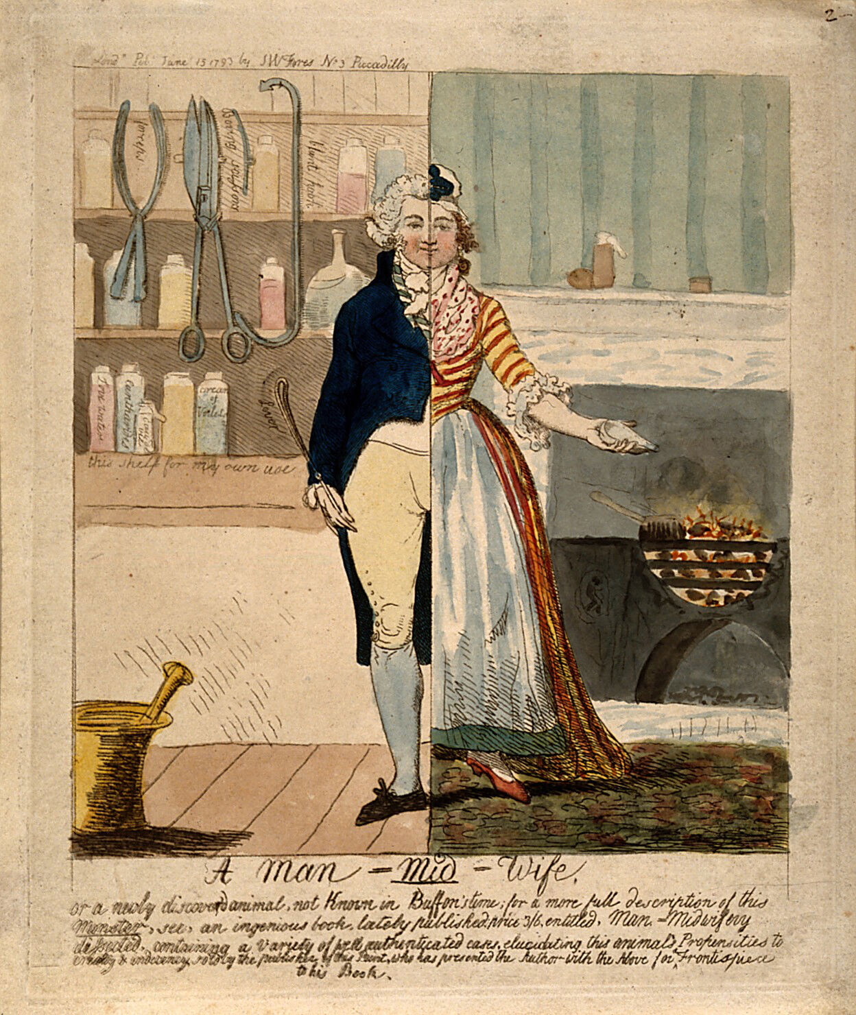
The history of the phantoms aligns with a significant change in the obstetrical practice, or rather, it can be understood as the outcome of this change. Until the seventeenth century, midwifery in the European continent was an occupation almost exclusively dominated by women. Occasionally, male surgeons (often referred to as accoucheurs) mainly assisted during abnormal births. But since the 1720s, their presence became more regular.[3] This is to be associated with the invention of obstetrical forceps, which led to the medicalization of the field. This evolution brewed rivalry between male midwives and female nurses and created a heated debate in the public discourse that was manifested in etchings such as “A man-midwife” (1793) (Fig. 2). For a long time, male midwives were viewed with suspicion.
As James Edgar (one of the most prominent obstetrical surgeons of the nineteenth century) notes, phantoms were brought into existence out of necessity.[4] Male midwives had limited access to deliveries until the eighteenth century. The presence of multiple men in the delivery room for training reasons would be deemed immoral. There were also social restrictions at play that further created obstacles to the teaching of obstetrics to male midwives. The presence of a man in such a private moment as birth, was deemed as disrespectful, not to the birthing woman but to her husband.[5] Furthermore, unlike other branches of medicine, obstetrical anatomy could not be taught by analyzing cadavers. Cadavers of parturient women were rare and difficult to access, and would have required the existence of big obstetric clinics, which did not emerge until the twentieth century.
Phantoms thus facilitated the education of these surgeons. However, they are not the earliest examples of models that represented the pregnant body. The first childbirth simulators date back to Renaissance Italy,[6] while archival sources reveal that the use of such models as tools for practical medical teaching preexisted in Germany and the Netherlands in the late seventeenth century. These were usually called “Phantome” or “fantomen”, and they consisted of simple, textile-and-leather models of the pelvis, uterus, and a doll-like fetus. They were used in midwife training, especially at universities and medical boards that sought to regulate midwifery.[7] By the eighteenth century, these models had a more complete form, depicting the abdomen and the thighs as well.

One of the first descriptions of obstetrical models was found in the medical handbook of Johan van Hoorn (1662-1724), a Dutch doctor who lived in Stockholm.[8] After finishing his studies in Leiden, Oxford, London, and Paris, Van Hoorn returned to Stockholm, where he taught obstetrics.[9] Apart from writing the first Swedish handbook for midwives, he also taught with the aid of a phantom: a doll with female genitalia, and a fetus made of leather.[10] This appears to be the first recorded use of an obstetric phantom for teaching.
At the beginning of the eighteenth century, we find Grégoire, a surgeon-accoucheur who offered private obstetric lessons with his son in Paris.[11] We do not have much information about his work or identity, but we know that doctors from all around Europe went to Paris for his lectures on obstetrics. Among the people who attended those lectures were William Smellie (1697-1763), and Jean-Georges Roeder (1726-1763), a prominent member of Göttinger University. Smellie and Roeder would create their own phantoms later on. Unfortunately, the Grégoire model did not survive. But we have a description of it in Smellie’s “Treatise on Midwifery” (1752):
“his machine was no other than a piece of basketwork, containing a real pelvis covered with black leather, upon which he could not clearly explain the difficulties that occur in turning children, proceeding from the contractions of the uterus, os internum, and os externum. And as for the forceps, he taught his pupils to introduce them at random, and pull with great force”.[12]
In 1756, Angélique Marguerite le Boursier du Coudray (1712-1794) presented “la machine”, her invention that helped her teach midwives in the rural parts of the kingdom on how to assist in deliveries. She insisted that the problem was that most sage-femmes (midwives) were illiterate, hence they could not access medical handbooks, and were unable to handle difficult cases. As a solution, she proposed a more practical education with the aid of the machine, along with an illustrated handbook. In 1759, King Louis XV issued her to travel around the countryside of the French Kingdom and provide midwifery education.[13]
Aside from being a midwife, Mme du Coudray was also a businesswoman, as she managed to commercialize her phantoms. Other people followed her footsteps, creating a whole market for obstetric models, and high demand for these objects persisted in France even after the Revolution.[14] René Levasseur (1747-1834), a surgeon-accoucheur from Mans, France, created another phantom in 1801, based on a human pelvis. A similar one created in 1820 was credited to Pierre-Louis Verdier. These surgeons also used cadavers of stillborn babies alongside the phantom for their presentations.[15]
During the nineteenth and twentieth centuries, the production of these mannequins continued and was also introduced to other continents. We have encountered obstetric dolls in Japan since the Edo period,[16] as in the United States from the eighteenth century onwards. While for Japan we have limited information on how they were developed (i.e., if they were imported from Europe, and under what circumstances), and who pioneered their use in teaching, we can assert with certainty that the first to use them in the United States was the surgeon and midwife William Shippen Jr. (1736-1808). Shippen traveled to England and Scotland and attended classes taught by William Hunter (1718-1783) and Colin Mackenzie (both of whom had studied under Smellie).[17] This network of doctors and their constant pursuit of knowledge, through travels and attending lectures given by experts, contributed to the expansion of the use of phantoms.
The Medical Gaze and the Fragmented Body
Having understood the context that allowed such an invention to materialize, we can now move to the social connotations of these objects. Hovering between scientific instruments and cultural artifacts, phantoms are now accredited with a dual nature. While their immediate function was pedagogical, enabling the simulation of childbirth, their materiality and form also render them aesthetic objects, with visual and material qualities that produce ideological meanings. They were not simply used to showcase medical knowledge: the way they were constructed also reflects biases, power structures, and social hierarchies of the time.
Phantom manufacturers depicted the parturient body in a fragmented way, with limbs and the upper body missing. Over time, this became the prevalent style in the production of these models. This choice reflected broader ideas about the perception of the female body that were dominant in the medical field at that time. Most phantoms were reduced to the female pelvis, lower abdomen, and uterus, with limbs and faces absent or only schematically rendered. This can act as an analogy to Michel Foucault’s medical gaze. In “The Birth of the Clinic”, Foucault uses this term to describe a mode of medical perception that abstracts the body into a series of signs and symptoms. This concept further signifies the reduction of the patient from a person with subjectivity to an object of knowledge.[18] Obstetric mannequins, in other words, visualize this medical reduction.
The choice of partial representation of the female body is not neutral; rather, it reflects a decision about which aspects or parts of the body are deemed worthy of representation. This is supported by the ideas that dominated medical thought at the time. Foucault, who studied this period in his “The Birth of the Clinic”, argues that towards the end of the eighteenth century, the “body” was created as the effect and object of medical examination.[19] By this, he refers to a shift in which the body, its function, and its significance came to be understood through medical observation and classification. The “medical gaze” transformed the body into an object that could be systematically studied and measured, affecting not only the way medical knowledge was produced, but also the ways in which the body was perceived and discussed, in both public and private discourse.[20]
The phantom, as argued above, can be viewed as a material manifestation of the medical gaze at an ideological level. By shifting the focus from the whole body to the uterus, it embodies a broader change in the medical perception of pregnant women. Within this framework, pregnant women were considered significant only for their ability to bear offspring, while phantoms were used to showcase primarily how this offspring would be delivered. In Foucauldian terms, the attention shifted from the patient (here, the female parturient body) to the symptom (pregnancy and more specifically, safeguarding the fetus). In other words, the phantom does not just simulate childbirth, but it stages the very reduction of the woman to a reproductive mechanism.
Moreover, the perception of the body was influenced by the popularization of dissections, which were conducted in public spaces.[21] Dissections moved the focus from the body as a whole to its fragmented parts.[22] While painting and sculpture promoted ideas about the perfect body type, anatomical drawings offered an idealized glimpse of the body’s interior. Separate organs or areas of the body started being discussed and illustrated individually. These depictions are examples of what Lorraine Daston and Peter Galison defined as “truth-to-nature.”[23] This meant not illustrating a specimen exactly as found but rather depicting an idealized version of it that embodied the “typical” structure of the organ.
This is evident in William Hunter’s seminal atlas “Anatomia uteri humani gravidi” (1774), where he depicted the human uterus in life-size proportions. At the same time, going through the handbooks or the notes of obstetrical surgeons, we see that they measured the “perfect” dimensions for the uterus.[24] This “golden rule” was applied in the making of obstetrical mannequins, as it was considered that this ratio was the most suitable for parturition, and as it would be beneficial to exercise upon this.[25] However, human bodies are diverse, and most pelvises are different. One can recall rachitic pelvis, a malformation of the pelvis due to lack of vitamin D, which was particularly common in Victorian England and rendered labor dangerous, if not impossible.[26] Hence, the phantom can be viewed as a materialization of a normative measurement that puts the bodies into categories.
This fragmented, idealized, and constructed body obliterates the natural experience of pregnancy. As knowledge moved away from the traditional practical experience of midwifery, the way birthing processes were understood became increasingly shaped by the medical training of male midwives. This was initially based on anatomical drawings, and the phantoms that came later, which were built according to this “golden rule” applied in these drawings. This enquiry for the perfect pelvis, the one with the best dimensions that would be most suitable for practical manipulation (in the literal meaning of the word), can be problematic. This, indirectly, led to a classification of the female body as perfect and not perfect based exclusively on its ability to deliver children. Barbara Duden, commenting on that, wrote that “the act of delivering a woman became the birth of a child.”[27] This is what the making of the phantoms implies: the focus is now on reproductive qualities and their outcome.
This fragmentary representation further supports the thesis that these objects of obstetric simulation did not focus on the experience of childbirth, but rather on the delivery of the fetus. More importantly, the absence of the limbs and the head on the mannequin testifies that this construction was not aiming at the reproduction of an actual birthing process, but it was simply a way to train on using forceps. Historical sources indicate that many women chose to give birth while kneeling or leaning on a chair or bed. Some midwives even carried birthing stools, specifically designed to make this process easier.[28] However, most phantoms depict the pregnant body lying down or in a small bent, a position that facilitates the gaze of the medical practitioner rather than improving the posture in order to alleviate the pain from this experience.
To sum up, midwifery, historically a domain occupied by women, was increasingly taken over by male physicians, who asserted epistemic authority through new technologies of visualization and simulation. Forceps and phantoms were central to this process. The invention of the latter allowed men to gain practical knowledge without attending actual births, thereby circumventing the need to rely on female midwives. The phantom was a material instrument, which marginalized the importance of female midwives. Scientific objects are never purely functional; they are also symbolic artifacts that have embedded social messages and that encode social hierarchies. The initial design of the forceps exemplifies that the comfort of the patient was not one of the main concerns; the obstetrical atlases testify to the canonization of the female anatomy. In the same way, obstetrical phantoms were mainly aiming at the safe delivery of the fetus, not the well-being of the mother. They are a direct side effect of the medical gaze and reflect the reduction of women to mere objects of knowledge.
Final Thoughts
Starting from an interesting and peculiar exhibit in a museum, we moved our focus towards medical perception and gender bias in the eighteenth and nineteenth centuries. By situating this mannequin within its historical, material, and visual contexts, this essay has sought to reveal the complex network of meanings embedded within such artifacts. Far from being neutral pedagogical devices, obstetric phantoms embody shifting medical practices, aesthetic ideals, and gendered power relations in Enlightenment and early modern Europe.
Obstetric phantoms emerged at a moment when male physicians increasingly entered a field traditionally dominated by female midwives. These devices allowed men to acquire hands-on experience without attending births, effectively displacing women’s embodied knowledge with visualized and mechanical forms of instruction. As Ludmilla Jordanova and Barbara Duden argue, this process involved a representational violence: the dissection, abstraction, and aestheticization of female bodies in medical imagery and objects were not neutral acts, but ways of exerting epistemic control.[29] The phantom materializes this exact shift. It is both a teaching tool and a symbol of the new gendered hierarchies in obstetric practice.
In the Boerhaave Museum today, the Salomon phantom invites new readings. No longer merely a tool for instructing future doctors, it has become a cultural artifact that reflects the tensions of its time. By examining its materials, form, and networks of production, we can uncover histories of gender, science, and representation that remain relevant to current debates about the body, technology, and knowledge. As Jordanova reminds us, the human body “can never be owned by a set of occupations”[30]: it remains a contested site of meaning. Obstetric phantoms remind us that medical knowledge is always produced through material forms.
Acknowledgements
I would like to express my gratitude to the Rijksmuseum Boerhaave in Leiden, for allowing me to conduct this research, and particularly to Ad Maas and Mieneke te Hennepe for their guidance and support.
Notes
[1] “Of Manikins and Machines: The Evolution of Obstetrical Phantoms,” Blog, Dittrick Medical History Center, October 15, 2013, https://artsci.case.edu/dittrick/2013/10/15/of-manikins-and-machines-the-evolution-of-obstetrical-phantoms/.
[2] Leonard F. Vernon, “A Brief Overview of How Male Medicine Co-Opted the Midwife’s Role in the Birth Process,” Open Journal of Nursing 5, no. 9 (2015): 758–64, https://doi.org/10.4236/ojn.2015.59079.
[3] Jean Donnison, Midwives and Medical Men : A History of the Struggle for the Control of Childbirth, (Routledge, 2023), https://doi.org/10.4324/9781003377948.
[4] James Clifton Edgar, “The Manikin in Teaching Obstetrics,” New York Medical Journal 52 (1890): 702.
[5] Ludmilla J. Jordanova, Nature Displayed: Gender, Science and Medicine 1760-1820 (Routledge, 2014), 152.
[6] Gaetano Giulio Zumbo (1656–1701) is considered one of the pioneers in this field. He created wax tableaux showing decomposition and disease as well as anatomical figures. His collaboration with surgeon Giovanni Maria Galli (c. 1690s) produced highly accurate anatomical waxes of the head and body. These are deemed as the earliest systematic anatomical wax models created with scientific intent.
[7] Such records are found mostly in Hamburg at the Collegium Medicum. For further information, see: Helen King, Midwifery, Obstetrics and the Rise of Gynaecology: The Uses of a Sixteenth-Century Compendium (Routledge, 2017).
[8] S. Kroes-Suverein, “De Vroedvrouw… Toen En Nu: Bevoegd En Bekwaam (The Midwife Then and Now; Authorized and Qualified).” Bilthoven: Catharina Schrader Stichting, 1998.
[9] Harry Owen, “Simulation in Obstetrics, Gynecology and Midwifery,” in Simulation in Healthcare Education: An Extensive History, ed. Harry Owen (Springer International Publishing, 2016), 71, https://doi.org/10.1007/978-3-319-26577-3_4.
[10] AO Lindfors, “II. Ueber Johan von Hoorn, Sein Leben Und Seine Werke,” Gynecologic and Obstetric Investigation 3, no. 1 (1896): 58-68.
[11] Kroes-Suverein, “De Vroedvrouw… Toen En Nu: Bevoegd En Bekwaam (The Midwife Then and Now; Authorized and Qualified).” and Owen, “Simulation in Obstetrics, Gynecology and Midwifery”. 75.
[12] William Smellie, Smellie’s Treatise on the Theory and Practice of Midwifery, vol. 79 (New Sydenham Society, 1876).
[13] Nina Rattner Gelbart, The King’s Midwife: A History and Mystery of Madame Du Coudray (University of California Press, 2023).
[14] Napoleon tried to reform healthcare and at the same time Baudelocque was appointed professor of midwifery at the newly created École de Santé.
[15] Harry Owen, ed., Simulation in Healthcare Education: An Extensive History (Springer International Publishing, 2016), https://doi.org/10.1007/978-3-319-26577-3_4. 6.
[16] The Dutch played an important role in bringing western medicine to Japan. Through Dutch translation of medical documents, Japanese doctors came in contact with Smellie’s anatomical atlas. RMF Van der Weiden et al., “The Continuing Influence of William Smellie (1697–1763) in Japan during the Early Meiji Period (1868–1880s),” Journal of Medical Biography 21, no. 3 (2013): 193–95. What is interesting about this object in the Japanese context is that they were not used only for teaching purposes but also for entertainment in the misenoto festivals (for more information see: “Morbid Anatomy: Japanese ‘Pregnancy Dolls,’ 19th Century,” Morbid Anatomy, May 12, 2009, https://morbidanatomy.blogspot.com/2009/05/japanese-pregnancy-dolls-19th-century.html.).
[17] Harry s, ed., Simulation in Healthcare Education: An Extensive History (Springer International Publishing, 2016), https://doi.org/10.1007/978-3-319-26577-3_4. 159.
[18] Michel Foucault, The Birth of the Clinic: An Archaeology of Medical Perception (Vintage Books, 1994). 107-122.
[19] Ibid. 130-150.
[20] Barbara Duden, The Woman Beneath the Skin: A Doctor’s Patients in Eighteenth-Century Germany (Harvard Univ. Press, 1998). 3.
[21] Renée Marlin-Bennet, et al., “Commodified Cadavers and the Political Economy of the Spectacle1,” International Political Sociology 4, no. 2 (2010): 159–77, https://doi.org/10.1111/j.1749-5687.2010.00098.x.
[22] Karl Figlio, “The Historiography of Scientific Medicine: An Invitation to the Human Sciences,” Comparative Studies in Society and History 19, no. 3 (1977): 277.
[23] Lorraine Daston and Peter Galison, Objectivity (Zone Books, 2007), JSTOR, https://doi.org/10.2307/j.ctv1c9hq4d.
[24] This was supported by Gottlieb Salomon and Petrus Camper to name a few examples in the Dutch context.
[25] Pam Lieske, “William Smellie’s Use of Obstetrical Machines and the Poor,” Studies in Eighteenth-Century Culture 29, no. 1 (2000): 65–86; Ludmilla J Jordanova, Nature Displayed: Gender, Science and Medicine 1760-1820 (Routledge, 2014). 101.
[26] Ann Louise Kibbie, Transfusion: Blood and Sympathy in the Nineteenth-Century Literary Imagination (University of Virginia Press, 2019).
[27] Barbara Duden, The Woman Beneath the Skin: A Doctor’s Patients in Eighteenth-Century Germany (Harvard Univ. Press, 1998). 18.
[28] Sarah Fox, Giving Birth in Eighteenth-Century England (University of London Press, 2022).
[29] Ludmilla J. Jordanova, Nature Displayed: Gender, Science and Medicine 1760-1820 (Routledge, 2014); Barbara Duden, The Woman Beneath the Skin: A Doctor’s Patients in Eighteenth-Century Germany (Harvard Univ. Press, 1998).
[30] Ludmilla J. Jordanova, Sexual Visions: Images of Gender in Science and Medicine Between the Eighteenth and Twentieth Centuries, Science and Literature (University of Wisconsin Press, 1989). 60.
About the author
Panagiota (Jota) Tsoutsa is an archeologist and a master’s student in Arts and Culture in the University of Leiden. Her research interests revolve around the intersection of arts and science through an ecological, posthuman, and gender-oriented lens.
Edited by Bianca Angelien Claveria and Lisa Vanderheyden